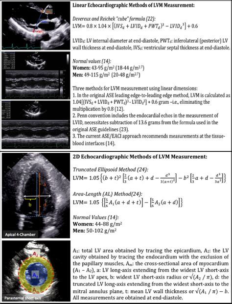
lv geometry | lvh measurements in echo lv geometry The first and most commonly used echocardiography method of LVM estimation is the linear method, which uses end-diastolic linear measurements of the interventricular . What is the application fee for LV Prasad Film and TV Academy? An application fee of INR 1500 is to be paid by the applicant in order to apply for the courses offered by LV Prasad Film and TV Academy.
0 · lvh measurements in echo
1 · lvh echo criteria wall thickness
2 · lvh criteria on echo
3 · lv wall thickness echo
4 · lv geometry calculator
5 · left ventricle wall thickness guidelines
6 · grading lvh on echo
7 · causes of lvh on echo
First in Fur. Fendi cultivates the most elevated craftsmanship, creating furs with its unique savoir-faire. This high attention to handcrafting the most extraordinary furs is a pursuit that has endured for over 90 years, producing a legacy of unprecedented invention and creativity in .Scalable from 5 kWh to 320 kWh. Maximum Flexibility for any Application with up to 64 ModulesConnected in Parallel. Compatible with Market Leading 1 and 3 Phase Inverters.Cobalt Free Lithium Iron Phosphate (LFP) Battery: Maximum Safety,Lifes.
Our LV calculator allows you to painlessly evaluate the left ventricular mass, left ventricular mass index (LVMI for the heart), and the relative wall thickness (RWT). Read on and discover all the details of our LV mass calculator and its variables: Definitions of abnormal LV .
Burton, R. Estimating body surface area from mass and height: theory and the . The first and most commonly used echocardiography method of LVM estimation is the linear method, which uses end-diastolic linear measurements of the interventricular . Our LV calculator allows you to painlessly evaluate the left ventricular mass, left ventricular mass index (LVMI for the heart), and the relative wall thickness (RWT). Read on and discover all the details of our LV mass calculator and its variables: Definitions of abnormal LV mass index; PWd normal range; and; IVSd in echo ️
The first and most commonly used echocardiography method of LVM estimation is the linear method, which uses end-diastolic linear measurements of the interventricular septum (IVSd), LV inferolateral wall thickness, and LV internal diameter derived from 2D-guided M-mode or direct 2D echocardiography. This method utilizes the Devereux and Reichek .
Our prospective cohort supports that abnormal LV geometry by echocardiography has a prognostic significance for incident stroke/CHD and all-cause mortality, implying that early detection and intervention of LV structural remodeling in rural China are urgently needed to prevent adverse outcomes. Greater left ventricular mass (LVM) and lower left ventricular (LV) systolic function, measured by echocardiography, are associated with excess adverse cardiovascular disease (CVD) events including coronary heart disease, 1 heart failure (HF), 2, 3, 4 stroke, 5 and both CVD and all‐cause mortality. 6, 7, 8, 9, 10 Lower LV systolic function .It has been recommended (9,10) to describe LV geometry as a function of LV mass and RWT. This leads to 4 categories: normal geometry, concentric remodeling (increased RWT), concentric hypertrophy (increased LV mass and RWT), and eccentric hypertrophy (increased LV .

The changes in left ventricular (LV) structure and geometry that evolve after myocardial injury or overload usually involve chamber dilation and/or hypertrophy. Such architectural remodeling can be classified as eccentric or concentric. Assessing LV systolic function in the presence of pathological remodeling is a common challenge in clinical practice. In addition to excellent prognostic abilities, GLS provides more accurate assessment of systolic function in conditions with increased LV mass or .
Increased left ventricular mass (LVM) is a strong independent predictor for adverse cardiovascular events, but conventional echocardiographic methods are limited by poor reproducibility and.
In this paper, we emphasize that the decrease in LVEF observed in HF is predominantly driven by changes in LV geometry and architecture. In support of this concept, we present data regarding biomarkers that can predict LV reverse remodelling and discuss their association with myocardial inflammation, fibrosis and cardiomyocyte stress. Based on established measures of LV mass and relative wall thickness (ratio of wall thickness to cavity diameter), four different LV geometric patterns were identified: normal geometry, concentric remodelling, concentric hypertrophy, and eccentric hypertrophy. Our LV calculator allows you to painlessly evaluate the left ventricular mass, left ventricular mass index (LVMI for the heart), and the relative wall thickness (RWT). Read on and discover all the details of our LV mass calculator and its variables: Definitions of abnormal LV mass index; PWd normal range; and; IVSd in echo ️ The first and most commonly used echocardiography method of LVM estimation is the linear method, which uses end-diastolic linear measurements of the interventricular septum (IVSd), LV inferolateral wall thickness, and LV internal diameter derived from 2D-guided M-mode or direct 2D echocardiography. This method utilizes the Devereux and Reichek .
Our prospective cohort supports that abnormal LV geometry by echocardiography has a prognostic significance for incident stroke/CHD and all-cause mortality, implying that early detection and intervention of LV structural remodeling in rural China are urgently needed to prevent adverse outcomes.
lvh measurements in echo
Greater left ventricular mass (LVM) and lower left ventricular (LV) systolic function, measured by echocardiography, are associated with excess adverse cardiovascular disease (CVD) events including coronary heart disease, 1 heart failure (HF), 2, 3, 4 stroke, 5 and both CVD and all‐cause mortality. 6, 7, 8, 9, 10 Lower LV systolic function .
It has been recommended (9,10) to describe LV geometry as a function of LV mass and RWT. This leads to 4 categories: normal geometry, concentric remodeling (increased RWT), concentric hypertrophy (increased LV mass and RWT), and eccentric hypertrophy (increased LV .The changes in left ventricular (LV) structure and geometry that evolve after myocardial injury or overload usually involve chamber dilation and/or hypertrophy. Such architectural remodeling can be classified as eccentric or concentric. Assessing LV systolic function in the presence of pathological remodeling is a common challenge in clinical practice. In addition to excellent prognostic abilities, GLS provides more accurate assessment of systolic function in conditions with increased LV mass or .
Increased left ventricular mass (LVM) is a strong independent predictor for adverse cardiovascular events, but conventional echocardiographic methods are limited by poor reproducibility and.
In this paper, we emphasize that the decrease in LVEF observed in HF is predominantly driven by changes in LV geometry and architecture. In support of this concept, we present data regarding biomarkers that can predict LV reverse remodelling and discuss their association with myocardial inflammation, fibrosis and cardiomyocyte stress.
lvh echo criteria wall thickness
Favorite Bistro. Claimed. Review. Share. 185 reviews #444 of 3,050 Restaurants in Las Vegas ££ - £££ American Bar Vegetarian Friendly. 3545 Las Vegas Blvd S # L13 L13, Las Vegas, NV 89109-8978 +1 702-844-4700 site Menu. Open now : 08:30 AM - 11:00 PM. Improve this listing. See all (356) Reserve a table. 2. Tue, 4/23. .
lv geometry|lvh measurements in echo


