lv volume overload | is lvh life threatening lv volume overload Left ventricular volume overload is the pathognomonic feature of chronic AR. The degree of volume overload is determined by the magnitude of the regurgitant flow, which is related to the . Eaton Magnum DS front access arc-resistant low voltage switchgear protects personnel from dangerous arcing faults by containing and redirecting the arc energy, with ratings up to 100 kA for 0.5 seconds.
0 · why is cardiac hypertrophy bad
1 · mild concentric lvh is dangerous
2 · life expectancy with lvh
3 · is lvh life threatening
4 · is hyperdynamic left ventricle dangerous
5 · dangers of left ventricular hypertrophy
6 · Lv overload or aspecific change
7 · Lv overload on ekg
E30 Tuning Stages Typical stage 1 mods often include: Panel air filter, Alloy wheels, Sports exhaust, Engine Tunes/Remapping, Lighter flywheel, Suspension upgrade (drop 23mm - 43 mm.). Typical stage 2 mods often include: high flow fuel injector, Ported and polished head, Power/Sport clutch, Fast road cam, fuel pump upgrades.
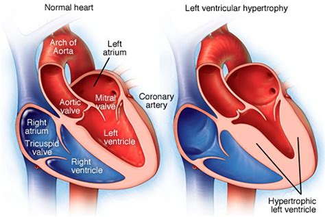
LV hypertrophy is a normal physiologic response to pressure and volume overload. Like any muscle, the heart grows bigger when it is forced to pump harder. In LV hypertrophy, the muscle fibers in the heart’s main pumping chamber enlarge and, over time, thicken.
Left ventricular volume overload is the pathognomonic feature of chronic AR. The degree of volume overload is determined by the magnitude of the regurgitant flow, which is related to the .
Left ventricular hypertrophy (LVH): Markedly increased LV voltages: huge precordial R and S waves that overlap with the adjacent leads (SV2 + RV6 >> 35 mm). R .A typical example of eccentric (volume-overload) geometry is that of mitral regurgitation (MR). The LV response to this volume overload consists of a progressive chamber enlargement with .Volume overload refers to the state of one of the chambers of the heart in which too large a volume of blood exists within it for it to function efficiently. Ventricular volume overload is .Left ventricular cavity dimensions and volumes are useful parameters to diagnose and monitor cardiac pathologies which lead to LV volume overload. 2-D echo/Doppler is the most .
why is cardiac hypertrophy bad
While deep q is very valuable in LV diastolic volume over load there are other useful ECG signs. Increased qrs amplitude (May be equally important like deep q . Both always go .
Aortic regurgitation (AR) is characterized by diastolic reflux of blood from the aorta into the left ventricle (LV). Acute AR typically causes severe pulmonary edema and .Left ventricular volume overload. This electrocardiographic changes occur in entities with increased diastolic wall stress of the left ventricle (Aortic regurgitation, mitral regurgitation, and .
LV hypertrophy is a normal physiologic response to pressure and volume overload. Like any muscle, the heart grows bigger when it is forced to pump harder. In LV hypertrophy, the muscle fibers in the heart’s main pumping chamber enlarge and, over time, thicken.Left ventricular volume overload is the pathognomonic feature of chronic AR. The degree of volume overload is determined by the magnitude of the regurgitant flow, which is related to the size of the regurgitant orifice, the aorta-ventricular pressure gradient, and the diastolic time.
mild concentric lvh is dangerous
Uncontrolled high blood pressure is the most common cause of left ventricular hypertrophy. Complications include irregular heart rhythms, called arrhythmias, and heart failure. Treatment of left ventricular hypertrophy depends on the cause. Treatment may include medications or surgery.
Left ventricular hypertrophy (LVH): Markedly increased LV voltages: huge precordial R and S waves that overlap with the adjacent leads (SV2 + RV6 >> 35 mm). R-wave peak time > 50 ms in V5-6 with associated QRS broadening. LV strain pattern with ST depression and T-wave inversions in I, aVL and V5-6.A typical example of eccentric (volume-overload) geometry is that of mitral regurgitation (MR). The LV response to this volume overload consists of a progressive chamber enlargement with characteristic changes in LV mass and RWT that depend, in part, on the severity and duration of the overload (41).Volume overload refers to the state of one of the chambers of the heart in which too large a volume of blood exists within it for it to function efficiently. Ventricular volume overload is approximately equivalent to an excessively high preload. It is a cause of cardiac failure.Left ventricular cavity dimensions and volumes are useful parameters to diagnose and monitor cardiac pathologies which lead to LV volume overload. 2-D echo/Doppler is the most commonly used imaging modality to assess LV volume overload in patients with valvular regurgitant disease.
life expectancy with lvh
While deep q is very valuable in LV diastolic volume over load there are other useful ECG signs. Increased qrs amplitude (May be equally important like deep q . Both always go together ) Aortic regurgitation (AR) is characterized by diastolic reflux of blood from the aorta into the left ventricle (LV). Acute AR typically causes severe pulmonary edema and hypotension and is a surgical emergency. Chronic severe AR causes combined LV .
Left ventricular volume overload. This electrocardiographic changes occur in entities with increased diastolic wall stress of the left ventricle (Aortic regurgitation, mitral regurgitation, and ventricular septal defect). LV hypertrophy is a normal physiologic response to pressure and volume overload. Like any muscle, the heart grows bigger when it is forced to pump harder. In LV hypertrophy, the muscle fibers in the heart’s main pumping chamber enlarge and, over time, thicken.
Left ventricular volume overload is the pathognomonic feature of chronic AR. The degree of volume overload is determined by the magnitude of the regurgitant flow, which is related to the size of the regurgitant orifice, the aorta-ventricular pressure gradient, and the diastolic time.
Uncontrolled high blood pressure is the most common cause of left ventricular hypertrophy. Complications include irregular heart rhythms, called arrhythmias, and heart failure. Treatment of left ventricular hypertrophy depends on the cause. Treatment may include medications or surgery. Left ventricular hypertrophy (LVH): Markedly increased LV voltages: huge precordial R and S waves that overlap with the adjacent leads (SV2 + RV6 >> 35 mm). R-wave peak time > 50 ms in V5-6 with associated QRS broadening. LV strain pattern with ST depression and T-wave inversions in I, aVL and V5-6.A typical example of eccentric (volume-overload) geometry is that of mitral regurgitation (MR). The LV response to this volume overload consists of a progressive chamber enlargement with characteristic changes in LV mass and RWT that depend, in part, on the severity and duration of the overload (41).Volume overload refers to the state of one of the chambers of the heart in which too large a volume of blood exists within it for it to function efficiently. Ventricular volume overload is approximately equivalent to an excessively high preload. It is a cause of cardiac failure.

Left ventricular cavity dimensions and volumes are useful parameters to diagnose and monitor cardiac pathologies which lead to LV volume overload. 2-D echo/Doppler is the most commonly used imaging modality to assess LV volume overload in patients with valvular regurgitant disease. While deep q is very valuable in LV diastolic volume over load there are other useful ECG signs. Increased qrs amplitude (May be equally important like deep q . Both always go together ) Aortic regurgitation (AR) is characterized by diastolic reflux of blood from the aorta into the left ventricle (LV). Acute AR typically causes severe pulmonary edema and hypotension and is a surgical emergency. Chronic severe AR causes combined LV .
is lvh life threatening

is hyperdynamic left ventricle dangerous
dangers of left ventricular hypertrophy
Labas preces par labām cenām. Akcijas preces no “Maxima Latvija” plašā sortimenta, kā arī Paldies kartes īpašos piedāvājumus.
lv volume overload|is lvh life threatening


